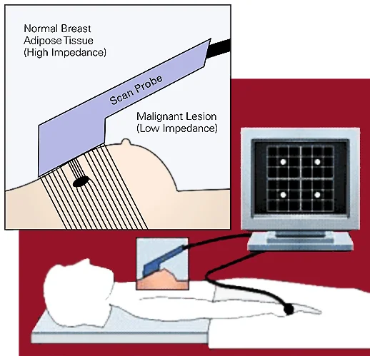
(Siemens, TS2000). Shown with reference electrode on right arm.
In EIT, a minute electrical current is injected into tissue and voltage measurements are taken at nearby electrodes across many signal frequencies, referenced to another electrode on the body.
Below: System diagram for a handheld cancer screening device using flexible electrode arrays. Waveform generation, sampling, voltage and current signal conditioning are performed in hardware, reducing required speed/bandwidth.
The measured voltage response is reconstructed into a 2-D impedance map, differentiating malignant tumors from high impedance healthy breast tissue. Visualization was performed offline by standard interpolation in Matlab, using spectral information from only the select frequencies generated by the device. Below: By sourcing and sinking current between different pairs of electrodes, voltages can be used to reconstruct images of tissue impedance/conductivity.
(Malvimuo, 1995. Bioelectromagnetism - Principles and Applications of Bioelectric and Biomagnetic Fields)
Contrasting conductivity of healthy vs. cancer tissue
Left: Conductivity plots (real and imaginary parts) of healthy adipose tissue vs. carcinoma. Note the large gap between red and blue traces, particularly at higher frequencies. Siemens TS2000 probes only in the ~1kHz range
(green band) leaving room for improvement in bandwidth with different sampling techniques and newer hardware. The goal was to build an inexpensive tool that takes advantage of higher spatial resolution and higher frequencies, by performing initial calculations in hardware before sampling. Right: Heatmaps showing 2mm tumor detection using standard vs. high density electrode arrays and 1 kHz vs. 100kHz signals (normalized, smoothed for visualization).
Tissue conductivities


Results: Standard resolution electrodes
Results: High density electrodes
Heatmaps generated by standard image interpolation in Matlab. Phantom ‘tumors’ were resolved with better SNR at high spatial resolution and higher frequencies, comparable to the best SNR from existing similar devices.
Data was taken from lab constructed breast phantoms, molded according to a standard recipe from the literature.
(Lazebnik, 2005. Tissue-mimicking phantom materials for narrowband and ultrawideband microwave applications).
(Left: Conductivity plots, Anderson, 2000. On Electrical Impedance Scanning - Principles and Simulations.)
Hardware


Above: System block diagram and PCB layout for a 4 channel current stimulation ‘blanking’ switchbox. During current injection, recording equipment must be automatically toggled to prevent saturation of precision amplifiers.
Below: 53x microscope photo of an electrode array designed to be implanted in a rat brain. After a successful proof of concept on dry skin, we used similar flexible electrode arrays and current stimulation to screen for breast cancer.
Electrode arrays and multiplexing PCB (4x 32 channel MUX). Hardware for neural studies or portable medical devices must be miniaturized and battery operated, with minimal wires due to assembly challenges and space constraints.